ã€introduction】
Nanomaterials and nanotechnology have received the attention of the scientific community in recent years, and their applications in various fields are becoming more and more extensive. Due to the special size effect of nanomaterials, the application of nanoparticles, nanotubes and various nanotechnology in biomedicine is booming and full of momentum. To date, many nanosystems have been developed as antibiotic alternatives for the treatment of bacterial infections. However, these advanced systems are limited due to their non-target aggregation and subsequent side effects.
[Introduction]
Recently, Dr. Wang Hao from the National Nanoscience Center and Associate Research Fellow Qiao Zengying (co-author) and others demonstrated pathologically-driven self-assembled nanostructures, which showed super accumulation at the target position due to the assembly-induced retention (AIR) effect. Retention ability. Inspired by this effect, this paper discusses a new antibacterial strategy—the “in-situ reassembly†strategy. With the aid of enzymes, the antibacterial active nanoparticles transform from spherical to fibrous structures, thus simultaneously in the bacterial infection site. Achieve long-term accumulation and enhance the efficacy of antibacterial. Related results were published in Adv. entitled "An "On-Site Transformation" Strategy for Treatment of Bacterial Infection". Mater. on.
[Graphic introduction]
Fig.1 Schematic diagram of self-assembly of CPC and principle of enzyme-induced morphological transformation
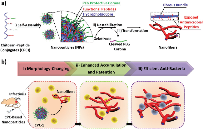
a) i) self-assembly of CPC into nanoparticles containing PEG shell; ii) cleavage of degradable peptide in the presence of gelatinase to strip the protective shell; iii) destruction of hydrophobic/hydrophilic equilibrium leading to chitosan Hydrogen bond interaction of the chain, spontaneously promotes self-assembly and reorganization of the fiber structure;
b) i) In the infective microenvironment, the CPC nanoparticles accumulated at the site of infection are cleaved by gelatinase produced by gelatinase-positive bacteria, causing in situ morphological transformation; ii) fibrous nanostructures are in situ in infected tissue Produced to allow nanomaterials to accumulate and their retention time to be extended; iii) Nanofibers with exposed antimicrobial peptides exhibit high antibacterial ability.
Figure 2 Variability characteristics of CPC-1
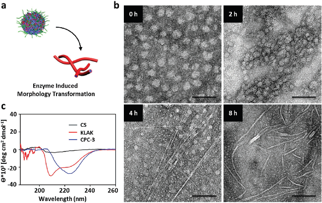
a) Schematic diagram of morphological transformation of CPC-1 under enzyme induction;
b) representative TEM image of each time period after immersing CPC-1 nanoparticles in gelatinase (10 μg/mL) tris buffer (pH 7.4) for a period of time; scale bar, 100 nm;
c) Chitosan (0.5 mg/mL), KLAK peptide and fiber CPC-3 (100×10-6 M, based on KLAK) in PB solution (10×10-3 M, pH 7.4) CD spectrum.
Figure 3 Synthesis scheme of CPCs
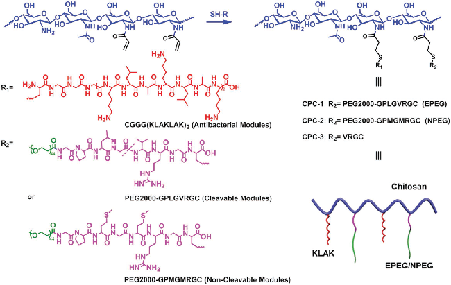
R1 represents the antimicrobial peptide KLAK; the conjugates are named CPC-1 and CPC-2; when R2 represents EPEG (gelatinase-cleaving peptide (GPLGVRGC) with PEG2000 terminus) and NPEG (control peptide with PEG2000 terminus (GPMGMRGC)) . The composition CPC-3 was used to simulate the gelatinase cleavage of CPC-1.
Figure 4 Interaction of in vitro CPCs with bacteria
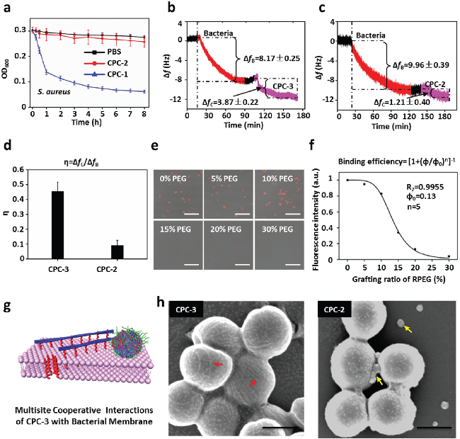
a) turbidity of the S. aureus solution treated with CPC-1 and CPC-2; a) turbidity of the S. aureus solution treated with CPC-1 and CPC-2;
b) Typical frequency change curve after injection of CPC-3 solution into a quartz crystal microbalance chamber. ΔfB and ΔfC represent the frequency changes of bacteria and CPC, respectively;
c) Typical curve of frequency change after injection of different solutions of CPC-2 into a quartz crystal microbalance chamber. ΔfB and ΔfC represent the frequency changes of bacteria and CPC, respectively;
d) the adsorption mass per unit of bacteria η = ΔfC / ΔfB = ΔmC / ΔmB, where ΔmC is the adsorption quality of CPC, ΔmB is the adsorption quality of the bacteria;
e) Fluorescent images of fluorescently labeled CPC-1 and S. aureus with different PEG graft ratios cultured in static culture for 1 hour. The fluorescence intensity of the bacteria is inversely proportional to the EPEG grafting rate of CPC-1. Scale bar, 10μm;
f) Hill index, n=5, indicating that the fiber CPC-1 has a strong multi-site binding to the bacterial surface;
g) Schematic diagram of the multi-site binding mode of CPC-3 to bacteria;
h) SEM images of S. aureus cultured with spherical CPC-2 and fiber CPC-3 for 1 hour. The yellow and red arrows represent nanoparticles and nanofibers, respectively. Scale bar, 500 nm.
Figure 5 Antibacterial activity of CPCs in vitro
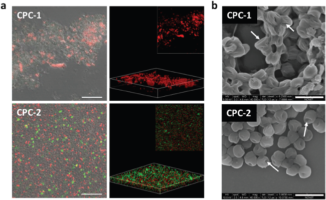
a) 2D and 3D confocal microscopy images of the killing potency of CPC-1 (top) and CPC-2 (bottom) in gelatinase-positive bacteria (Gram-positive bacteria, Staphylococcus aureus). Scale bar, 10μm;
b) The morphology of S. aureus cultured for 6-8 hours with CPC-1 (top) and CPC-2 (bottom) (300 x 10-6 M), with arrows indicating damage, collapse and fusion of the bacterial membrane. Scale bar, 2μm.
Figure 6. Accumulation, retention and antibacterial activity of CPCs in vivo

a) In vivo near-infrared fluorescence imaging of S. aureus after injection of 1 PBS, 2CPC-2, 3CPC-1. A PBS buffer solution (pH 7.4) was used as a control. A white circle indicates the site of infection;
b) Average fluorescence signal at different sites of infection at different times. Red arrows indicate injections on day 0, day 1, day 2 and day 3;
c) CLSM images of infected tissue sections of mice injected with PBS, CPC-2 and CPC-1. Arrows indicate fibrous fluorescent signals. The insertion map is a high magnification image of the white arrow area. Scale bar, 100×10-6m;
d) TEM image of nanofibers formed at the site of infection of S. aureus after injection of 8 days of CPC-1 in the tail vein compared to the site of infection treated with CPC-2 (insert in the lower panel). Arrows indicate the fiber structure. Scale bar, 1 μm. The corresponding energy dispersion map acquired in the region of interest is marked by a red star in Figure d (top insert);
e) in vitro near-infrared fluorescence images of tissues and major organs post-treated by PBS, CPC-2 and CPC-1 for 8 days;
f) Average fluorescence signal of major organs after 8 days of tail vein injection. The average fluorescence signal value is expressed as mean ± S. D (N=6). The asterisk (**) indicates the statistical significance of **P < 0.01; g) the survival of S. aureus in the infected area was quantified on day 5. The insert map shows bacterial colonies of infected tissues of mice injected with PBS, CPC-1 and CPC-2. Error bars were determined from 5 mice per group.
ã€summary】
Researcher Wang Hao et al. developed a CPCs containing gelatinase cleavage EPEG and antibacterial peptide KLAK, which self-assembled into nanoparticles during the reaction. The recombination of CPC-1 significantly improved the interaction between nanomaterials and bacteria through a multi-site synergistic electrostatic binding model between exposed KLAK and bacterial membranes, and the antibacterial efficacy was significantly enhanced. Compared to the invariant CPC-2, the transformable CPC-1 has a longer retention time and better in vivo antibacterial properties.
Adult TPE love dolls can be very beneficial when it comes to becoming a pro in the bedroom.
Our world-class collection of silicon wives with big breast goes long way to satisfy your sexual desires and make masturbation an inventive and fun process.

All of these TPE love dolls with big breast are made with extreme care to provide you with highest satisfaction to the customers.

Men from all over the world have experienced intense orgasms from these big breast TPE love dolls which also features an open-mouth in addition to the love tunnels front side and back side.

Big Boobs Sex Dolls,Bbw Big Boobs Sex Dolls,Bbw Women,Realistic Bbw Big Boobs Sex Dolls
Dongguan Chenkuang Biological Technology Co., Ltd , https://www.cksexdoll.com